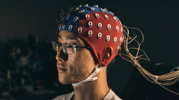
Every brain-computer interface makes the same bargain: the closer your electrodes sit to the neurons you want to read, the better the signal—and the higher the clinical risk. EEG records from the scalp with no surgery at all, electrocorticography (ECoG) places electrodes on the brain's exposed surface through a surgical opening, and intracortical microelectrodes are implanted directly into brain tissue during neurosurgery. Understanding where each modality falls on that spectrum is the key to evaluating any BCI claim, whether the application is consumer neurofeedback or prosthetic control in a clinical trial.
Understanding Brain-Computer Interfaces: The Core Trade-Off
A brain-computer interface is a system that records neural activity and converts it into commands for an external device. As one review of BCIs in medicine recounts, researchers have progressed from early electroencephalography-based spelling demonstrations and single-neuron device control to increasingly complex control of cursors, robotic arms, prostheses, and wheelchairs, with proposed uses extending to stroke rehabilitation and even augmenting surgeon performance.
What separates the modalities is not the goal but the recording location. As Neuromind's technical overview of BCI systems puts it, different recording techniques offer "different compromises between precision, accessibility and clinical risk." That single sentence describes the entire decision space: each step deeper into the skull buys precision while costing accessibility and adding risk.